Medical Devices and Materials Engineering SectionCognitive Neuroscience
Approaching human brain function across multiple spatial scales using laminar fMRI
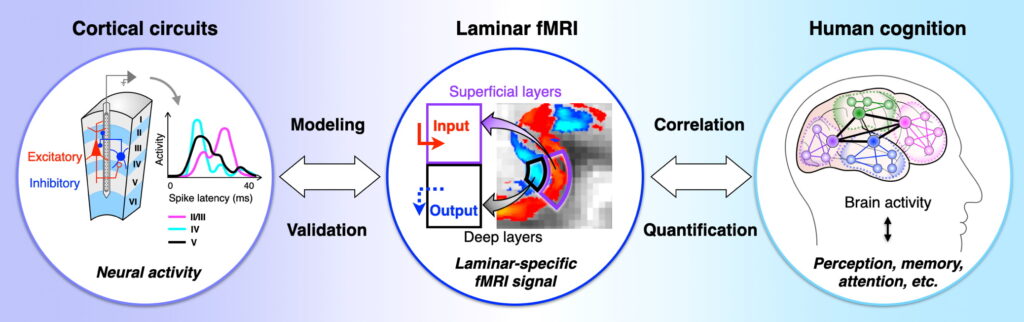
Recently devised in vivo ultra-high-field (UHF), high-resolution (submillimeter-level) functional magnetic resonance imaging (fMRI) has begun to directly reveal laminar-specific brain activity in the human brain. The current spatial resolution capability of UHF MRI in the human brain at 7T stands at approximately 0.3 mm isotropic resolution for structural images and approximately 0.8 mm resolution for functional images. This noninvasive imaging method, known as laminar bridges the gap between findings from laboratory animals and humans. In this project, our laminar investigation was made possible by using the new method VASO in place of the conventional gradient-echo BOLD contrast. Combining laminar fMRI with invasive neurophysiological measures in nonhuman primates will help us to establish a modeling-validation loop to identify potential relationships between neural activity and the fMRI signal. Moreover, the higher spatial resolution of laminar fMRI enhances our capacity to resolve directional connectivity in brain networks by adding the third dimension of the cortex.
Faculty
Medical Devices and Materials Engineering Section Cognitive Neuroscience